Publications
2024
-
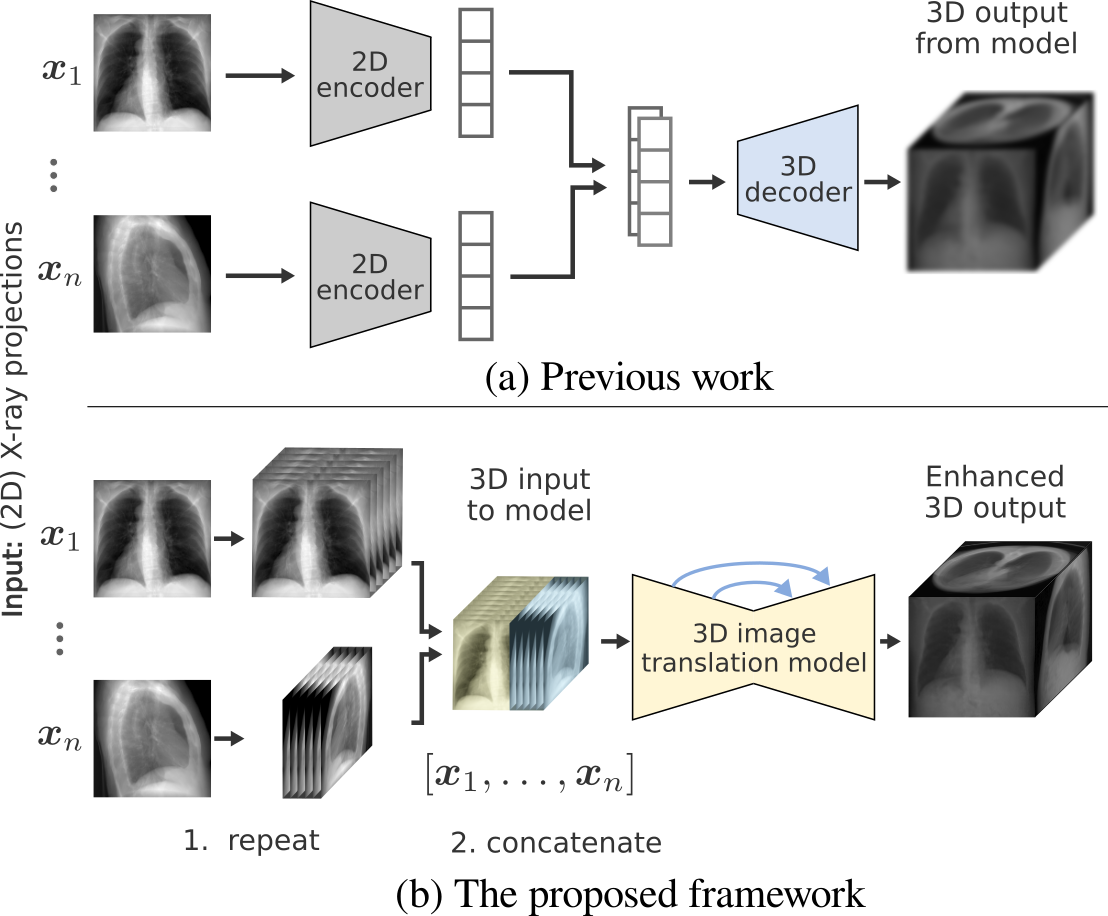 Repeat and Concatenate: 2D to 3D Image Translation with 3D to 3D Generative ModelingAbril Corona-Figueroa, Hubert P. H. Shum, and Chris G. WillcocksIn Proceedings of the IEEE/CVF Conference on Computer Vision and Pattern Recognition (CVPR) Workshops, Jun 2024
This paper investigates a 2D to 3D image translation method with a straightforward technique enabling correlated 2D X-ray to 3D CT-like reconstruction. We observe that existing approaches which integrate information across multiple 2D views in the latent space lose valuable signal information during latent encoding. Instead we simply repeat and concatenate the 2D views into higher-channel 3D volumes and approach the 3D reconstruction challenge as a straightforward 3D to 3D generative modeling problem sidestepping several complex modeling issues. This method enables the reconstructed 3D volume to retain valuable information from the 2D inputs which are passed between channel states in a Swin UNETR backbone. Our approach applies neural optimal transport which is fast and stable to train effectively integrating signal information across multiple views without the requirement for precise alignment; it produces non-collapsed reconstructions that are highly faithful to the 2D views even after limited training. We demonstrate correlated results both qualitatively and quantitatively having trained our model on a single dataset and evaluated its generalization ability across six datasets including out-of-distribution samples.
@inproceedings{Corona-Figueroa_2024_CVPR, author = {Corona-Figueroa, Abril and Shum, Hubert P. H. and Willcocks, Chris G.}, booktitle = {Proceedings of the IEEE/CVF Conference on Computer Vision and Pattern Recognition (CVPR) Workshops}, series = {CVPRW '24 DCA in MI}, title = {Repeat and Concatenate: 2D to 3D Image Translation with 3D to 3D Generative Modeling}, year = {2024}, month = jun, publisher = {IEEE/CVF}, location = {Seattle, USA}, } -
 2D to 3D Translation with Medical Image ApplicationsAbril Corona-FigueroaJun 2024Doctoral thesis
To translate between medical images of different modalities, one option is to rely on novel deep learning approaches within the broad task of image-to-image translation. Over the past decade, this problem has spawned many computer vision subtasks, including image inpainting, domain adaptation, semantic segmentation, super-resolution, among others. However, adapting or designing deep learning frameworks for real-world data, such as medical imaging, imposes additional considerations in terms of data availability, structure, and topology. In particular, 2D to 3D medical image translation entails further complexities: 1. Lack of unpaired datasets makes learning direct mappings difficult, thus precise transformations cannot be guaranteed. 2. Dimensionality mismatch calls for a generative modeling formulation where issues such as poor spatial alignment and impractical data representations can lead to inaccurate translations. 3. Hallucination can prove fatal in medical contexts. Unlike in a "natural image" setting, which can focus on aesthetics or creativity, medical imaging relies on the precise representation of anatomical structures. The thesis makes advancements in such areas and works towards having positive outcomes in applications like 3D CT reconstruction from 2D X-rays, involving lower radiation doses for patients, cost savings, and increased accessibility. Specifically, the research work presented in this thesis explores different deep learning approaches for the task of 2D to 3D translation, considering necessary data domain and technical aspects. Our proposed methods generally outperform competitive models and introduce novel ideas with potential use in other frameworks or relevant applications. These methods have been implemented in PyTorch and are open source for the research community. This work aims to contribute to the problem of 2D to 3D image translation using deep learning. To complement existing methods for CT reconstruction from X-rays, which primarily focus on adapting architectures to medical images, we concentrate on studying the data structure and making well-founded design decisions to effectively solve modeling issues and achieve state-of-the-art results. This thesis is also intended to be accessible to researchers without a computer science background, promoting multidisciplinary collaboration with clinicians and biomedical experts.
2023
-
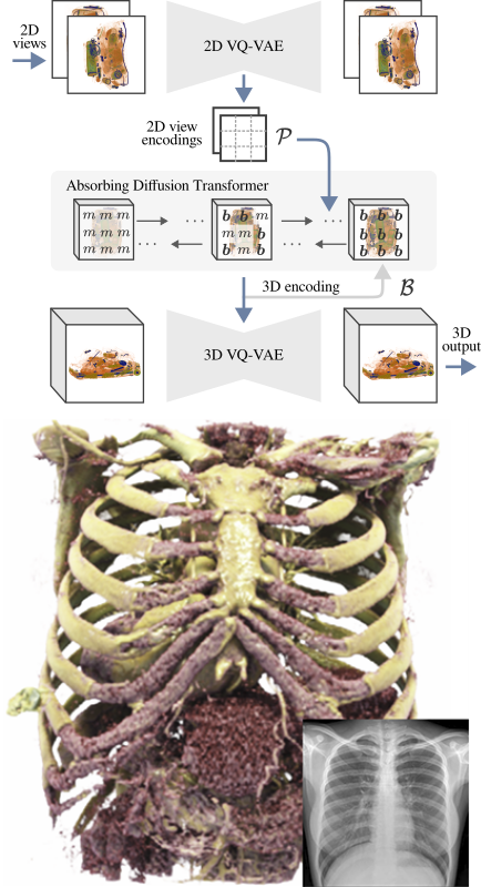 Unaligned 2D to 3D Translation with Conditional Vector-Quantized Code Diffusion using TransformersAbril Corona-Figueroa, Sam Bond-Taylor, Neelanjan Bhowmik, Yona Falinie A. Gaus, Toby P. Breckon, Hubert P. H. Shum, and Chris G. WillcocksIn Proceedings of the 2023 IEEE/CVF International Conference on Computer Vision, Oct 2023
Generating 3D images of complex objects conditionally from a few 2D views is a difficult synthesis problem, compounded by issues such as domain gap and geometric misalignment. For instance, a unified framework such as Generative Adversarial Networks cannot achieve this unless they explicitly define both a domain-invariant and geometric-invariant joint latent distribution, whereas Neural Radiance Fields are generally unable to handle both issues as they optimize at the pixel level. By contrast, we propose a simple and novel 2D to 3D synthesis approach based on conditional diffusion with vector-quantized codes. Operating in an information-rich code space enables high-resolution 3D synthesis via full-coverage attention across the views. Specifically, we generate the 3D codes (e.g. for CT images) conditional on previously generated 3D codes and the entire codebook of two 2D views (e.g. 2D X-rays). Qualitative and quantitative results demonstrate state-of-the-art performance over specialized methods across varied evaluation criteria, including fidelity metrics such as density, coverage, and distortion metrics for two complex volumetric imagery datasets from in real-world scenarios.
@inproceedings{coronafigueroaa23unaligned,
  author = {Corona-Figueroa, Abril and Bond-Taylor, Sam and Bhowmik, Neelanjan and Gaus, Yona Falinie A. and Breckon, Toby P. and Shum, Hubert P. H. and Willcocks, Chris G.},
  booktitle = {Proceedings of the 2023 IEEE/CVF International Conference on Computer Vision},
  series = {ICCV '23},
  title = {Unaligned 2D to 3D Translation with Conditional Vector-Quantized Code Diffusion using Transformers},
  year = {2023},
  month = oct,
  publisher = {IEEE/CVF},
  location = {Paris, France},
}
2022
-
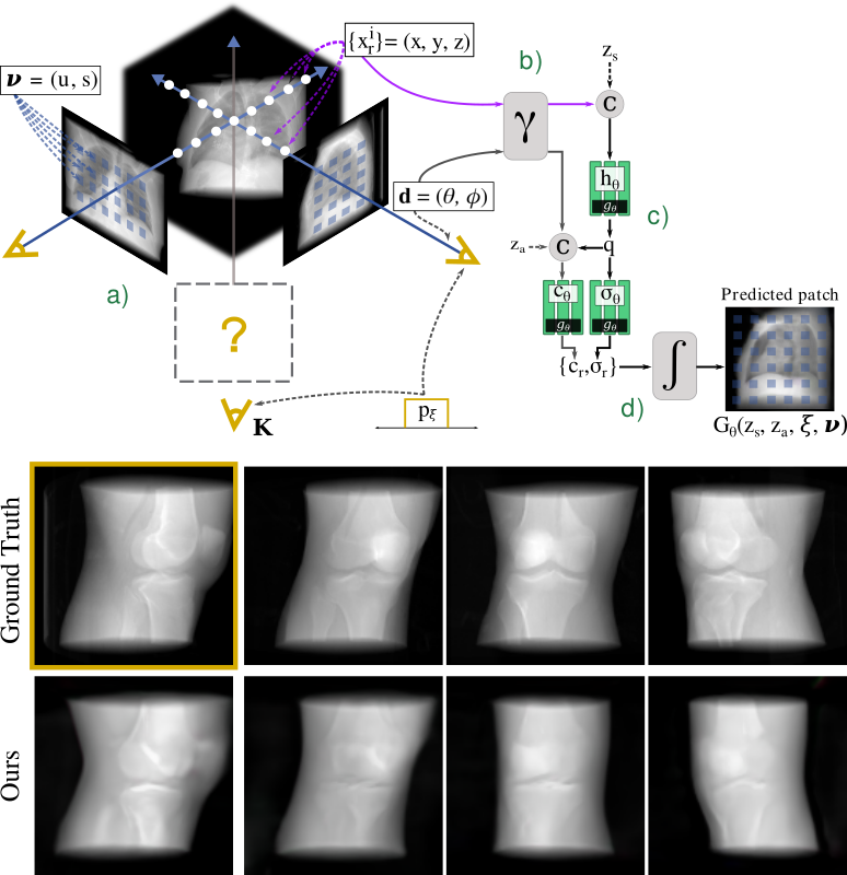 MedNeRF: Medical Neural Radiance Fields for Reconstructing 3D-aware CT-Projections from a Single X-rayAbril Corona-Figueroa, Jonathan Frawley, Sam Bond-Taylor, Sarath Bethapudi, Hubert P. H. Shum, and Chris G. WillcocksIn 2022 44th Annual International Conference of the IEEE Engineering in Medicine & Biology Society (EMBC), Oct 2022
Computed tomography (CT) is an effective med-ical imaging modality, widely used in the field of clinical medicine for the diagnosis of various pathologies. Advances in Multidetector CT imaging technology have enabled additional functionalities, including generation of thin slice multi planar cross-sectional body imaging and 3D reconstructions. However, this involves patients being exposed to a considerable dose of ionising radiation. Excessive ionising radiation can lead to deterministic and harmful effects on the body. This paper proposes a Deep Learning model that learns to reconstruct CT projections from a few or even a single-view X-ray. This is based on a novel architecture that builds from neural radiance fields, which learns a continuous representation of CT scans by disentangling the shape and volumetric depth of surface and internal anatomical structures from 2D images. Our model is trained on chest and knee datasets, and we demonstrate qual-itative and quantitative high-fidelity renderings and compare our approach to other recent radiance field-based methods. Our code and link to our datasets are available at https://qithub.com/abrilcf/mednerf Clinical relevance- Our model is able to infer the anatomical 3D structure from a few or a single-view X-ray showing future potential for reduced ionising radiation exposure during the imaging process.
@inproceedings{9871757,
  author = {Corona-Figueroa, Abril and Frawley, Jonathan and Bond-Taylor, Sam and Bethapudi, Sarath and Shum, Hubert P. H. and Willcocks, Chris G.},
  booktitle = {2022 44th Annual International Conference of the IEEE Engineering in Medicine & Biology Society (EMBC)},
  title = {MedNeRF: Medical Neural Radiance Fields for Reconstructing 3D-aware CT-Projections from a Single X-ray},
  year = {2022},
  volume = {},
  number = {},
  pages = {3843-3848},
  doi = {10.1109/EMBC48229.2022.9871757},
  dimensions = {false},
}
2019
-
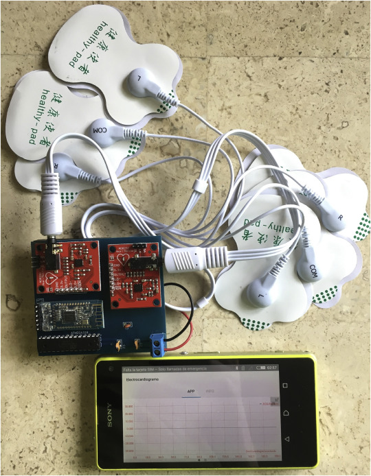 A portable prototype for diagnosing fetal arrhythmiaAbril Corona-FigueroaInformatics in Medicine Unlocked, Oct 2019
Expert knowledge and proper techniques are necessary for diagnosing and treating fetal cardiac disease. Fetal arrhythmia represents an important part of fetal cardiac disease as it conveys abnormalities in the electrophysiological state of the heart. Arrhythmias can be caused by rate or rhythm alterations or by abnormalities in QRS or QT intervals. Fetal echocardiography has been the gold standard for pediatric cardiologists since it assesses multiple aspects of fetal cardiovascular pathology. In parallel, artificial intelligence techniques are being designed to provide additional support to experts in the medical field in terms of detection and accuracy. The present manuscript describes the development of a prototype meant to be an auxiliary for the detection of fetal arrhythmias by analyzing the fetal heart rate (FHR) and its variability. It consists of a portable electrocardiograph and a mobile application that, together, extract the fetal electrocardiogram for its analysis to finally render a first diagnosis. Conducted tests with synthetic signals based on clinical fetal arrhythmia provided a detection rate of 88.88%. Results from tests with pregnant women may indicate that the proposed prototype can be used to render a first diagnosis without entirely depending on expert assistance. This prototype might be helpful to perform routine check-ups in the second or third trimester in pathological pregnancies associated with high risk of onset/progression of fetal arrhythmias. In addition, medical practitioners could use it to corroborate a given diagnosis or in scenarios where there is an insufficiency of human experts to attend a high population of patients.
@article{CORONAFIGUEROA2019100268,
  title = {A portable prototype for diagnosing fetal arrhythmia},
  journal = {Informatics in Medicine Unlocked},
  volume = {17},
  pages = {100268},
  year = {2019},
  issn = {2352-9148},
  doi = {https://doi.org/10.1016/j.imu.2019.100268},
  url = {https://www.sciencedirect.com/science/article/pii/S2352914819301893},
  author = {Corona-Figueroa, Abril},
}
Conference Abstracts
2026
-
 Generalizable Adverse Safety Event Detection with Locally Deployable Language ModelsAbril Corona-Figueroa, Nathan Bahr, Steven Bedrick, Matthew Hansen, Xiaoqian Jiang, and Jeanne-Marie GuiseIn Mobilizing Computable Biomedical Knowledge, Oct 2026Abstract will be published in the Learning Health Systems Journal
Background: Adverse safety events (ASEs) are a leading cause of death in the United States. Pediatric ASEs are of special interest because of their high acuity; however, their detection is more difficult due their low prevalence. Moreover, the adjudication of errors requires expert knowledge to accurately discern case context and documented procedures in emergency medical service charts. These limit the scalability of manual review, presenting the opportunity to develop novel approaches that can help with the systematic detection of ASEs while preserving clinical knowledge required for their identification. Methods: We previously developed a framework for the detection of ASEs in pediatric cardiac arrest emergencies. The framework consisted of large language models (LLMs) for information extraction, guided by graph reasoning based on ontologies derived by experts for both medications and procedures. In this work, we evaluate and analyze its implementation feasibility and generalization across various locally deployed LLMs. Using the same knowledge representation, prompts, and processing strategy across models, we examine the relationship between ASE detection performance, model parameter count, inference time, and model reasoning capabilities. Results show insights into the potential tradeoffs between detection performance and computational efficiency, which are informative for prior implementation assessment within institution environments. Results and Discussion: Contrary to expectation, across evaluated models, larger LLMs were not necessarily associated with better detection performance of ASEs. For example, DeepSeek-R1 32B and its 8B version achieved similar performance. On the other hand, several smaller models achieved comparable performance in similar inference times, indicating that the incorporation of expert knowledge into the graph reasoning framework reduces the dependence on model scale. Models that have reasoning capabilities were able to achieve higher detection performance but required longer inference times. Overall, we observed that short and clear prompts are effective across models as they allow more model capacity to be used for context reasoning. Furthermore, chunking strategies provided a controlled inference setting regardless of context windows and reasoning capacity. Conclusion: Our work shows preliminary data that the proposed ASE detection framework is agnostic to LLM selection, further enabling its use when limited computational resources are available. The combination of expert knowledge as graphs, efficient prompting and a chunking strategy greatly improves the portability of the framework and overall reproducibility of results, enabling its potential implementation for safety surveillance.
-
 Urban and Rural Differences in Pediatric Mortality: A National Analysis of EMS RecordsAbril Corona-Figueroa, Trang Huynh, Matthew Hansen, and Jeanne-Marie GuiseIn AcademyHealth Annual Research Meeting, Jun 2026Accepted conference abstract
Research Objective: Recent evidence indicates that children living in rural areas experience higher mortality than those in urban settings, often attributed to unintentional injuries, though the underlying causes are unclear. This study aimed to examine pediatric mortality differences among EMS patients by scene location. Study Design: We conducted a retrospective analysis of pediatric (<18 years) 9-1-1 encounters from the 2018-2023 ESO Data Collaborative research datasets. We standardized records and compared mortality across urbanicity classifications to identify vulnerable populations and geographic differences. Population Studied: Over the five-year period, there were 3,756,061 EMS encounters for children aged 0–17 years; 3,119,180 urban encounters (83%), 527,352 rural (14%), and 96,809 (3%) super-rural. Principal Findings: Super-rural pediatric mortality was nearly twice that of urban areas (RR = 1.97, 95% CI 1.79–2.15) and 37% higher than rural areas (RR = 1.37, 95% CI 1.23–1.51), despite most deaths occurring in urban settings (2.6 per 1,000 EMS encounters vs. 3.7 rural and 5 super-rural per 1,000). Mortality was higher in males than females (RR = 1.59, 95% CI 1.53–1.66). Although adolescents (13–17 years) accounted for the largest share of encounters (41.8%), mortality was highest among neonates (RR = 5.13, 95% CI 4.82–5.46) and infants (RR = 3.24, 95% CI 2.93–3.58), with 17-year-olds showing lower rates (3 per 1,000). Among 10,508 pediatric deaths, cardiac arrest was the most common primary impression (42.5%), with infant asphyxia responsible for the largest number of cardiac-arrest deaths (n = 304) and trauma predominating among adolescents. In deaths with documented timing (45%), pre-EMS deaths were proportionally higher in rural (21%) and super-rural areas (17%) than in urban regions (3.6%), whereas urban deaths more often occurred in the ED (23%). Neonates and infants comprised the largest pre-EMS death group across geographies, followed by 17-year-olds. Trauma was the leading recorded etiology in all settings; exceeding urban levels by at least 15% in rural and super-rural regions, followed by presumed cardiac and respiratory/asphyxia causes. Firearm injuries predominated in urban areas, motor vehicle crashes in rural and super-rural regions, and intentional self-harm was least frequent in super-rural areas. Median EMS response times were shortest in urban areas (6.6 min, IQR 4.6–9.5), slightly longer in rural areas (7.2 min, IQR 4.7–11.2), and in super-rural regions (7 min, IQR 4.5–13). Conclusions: Children in rural and super-rural areas had higher mortality across age groups and etiologies, with neonates and infants at greatest risk. Implication for Policy or Practice Neonates and infants are at highest risk for mortality across settings. They are also groups with unique equipment sizing and medication dosing needs. These findings suggest that efforts to ensure that rural communities and services have the resources and support to be prepared for pediatric emergencies are justified.
-
 Development and pilot testing of a linear cognitive app to support EMS teams in neonatal and pediatric cardiac arrest resuscitationsAbril Corona-Figueroa, and Jeanne-Marie GuiseIn Pediatric Academic Societies Meeting, Apr 2026Accepted conference abstract
Background: Pediatric cardiac arrest is a leading cause of child mortality. Most events occur out-of-hospital, where Emergency Medical Services (EMS) provide critical care. Because neonatal and pediatric cardiac arrests are relatively rare, EMS providers have limited exposure and may face higher cognitive demands. Prior studies found that non-linear cognitive aids were associated with delays in cardiac arrest care. Objective: To develop and test the feasibility of a linear cognitive app (RESCUER) intended to support EMS personnel in responding to neonatal and pediatric out-of-hospital cardiac arrest (POHCA). Design/Methods: Development and pilot testing of a linear cognitive app that combines NRP and PALS step-by-step algorithmic guidance with age and weight-based pediatric dosing of medications and sizing of equipment. Its linear design provides sequential real-time instructions during resuscitations. We also developed voice-guidance that functions much like AED voice prompts, to allow all team members to stay on track. We conducted preliminary feasibility testing with EMS teams in preparation for a larger planned RCT. Results: Preliminary tests with rural and urban EMS teams found that users were able to easily understand how to use the app with very little orientation (<5 minutes). Users were also able to use the app in simulated cardiac arrest settings without major disruption. However, in one instance, the person holding the app got so enthralled with the simulation that they forgot to start the app for quite a while. By the time they started it, given its linear nature, the instructions were behind the team. While one test among a team consisting of highly experienced EMS trainers found the linear nature frustrating stating that they were thinking many steps ahead, a rural team consisting of newly minted EMTs reported that the app was highly valuable. We also identified one save where a team proceeded with the wrong treatments until the app gave instructions for the correct steps. Voice commands also received mixed results with experienced teams finding it distracting and less experienced rural teams finding it calming in the setting of events which they find highly stressful. Conclusions: Findings from our pilot suggest that using an app to guide clinical care for POHCA, is likely feasible in a variety of settings. It also provided interesting hypotheses regarding the role of experience with linear design and voice prompts. We have started a randomized control trial to evaluate the app in rural and urban EMS practices (NCT06768099 clinical trials.gov).